
Model for Hematoma
To design a pelvic model to teach healthcare professionals how to identify, assess, and treat hematomas

The Need
Ten years ago, when people needed a cardiac catheterization procedure, the most common access point was through blood vessels in the femoral region. Following the procedure, nurses examined the patient for complications. One of the most common complications is a hematoma that develops in the femoral area. A hematoma can cause excruciating pain and even become life- threatening. In recent years, catheters have become thinner and more flexible, meaning they can be inserted through arteries and veins in the wrist. This procedure has become the standard for cardiac catheterization; however, in certain situations, catheterization through the femoral region is necessary. The issue is that nurses now have not been taught to identify and treat hematomas in the femoral region after a catheter procedure. To address this issue, we are creating a pelvic model to teach healthcare professionals how to identify, assess, and treat hematomas
The Solution
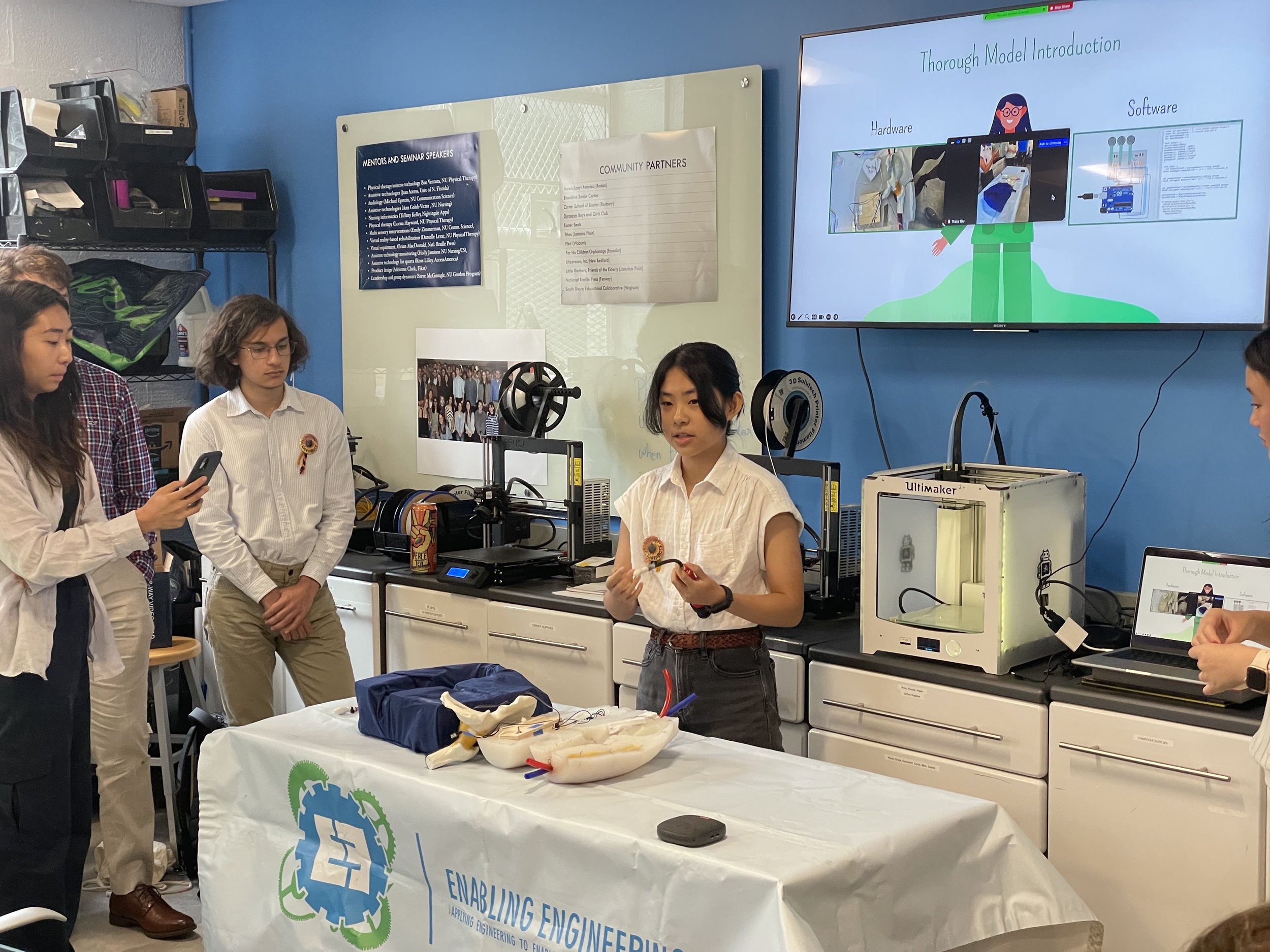
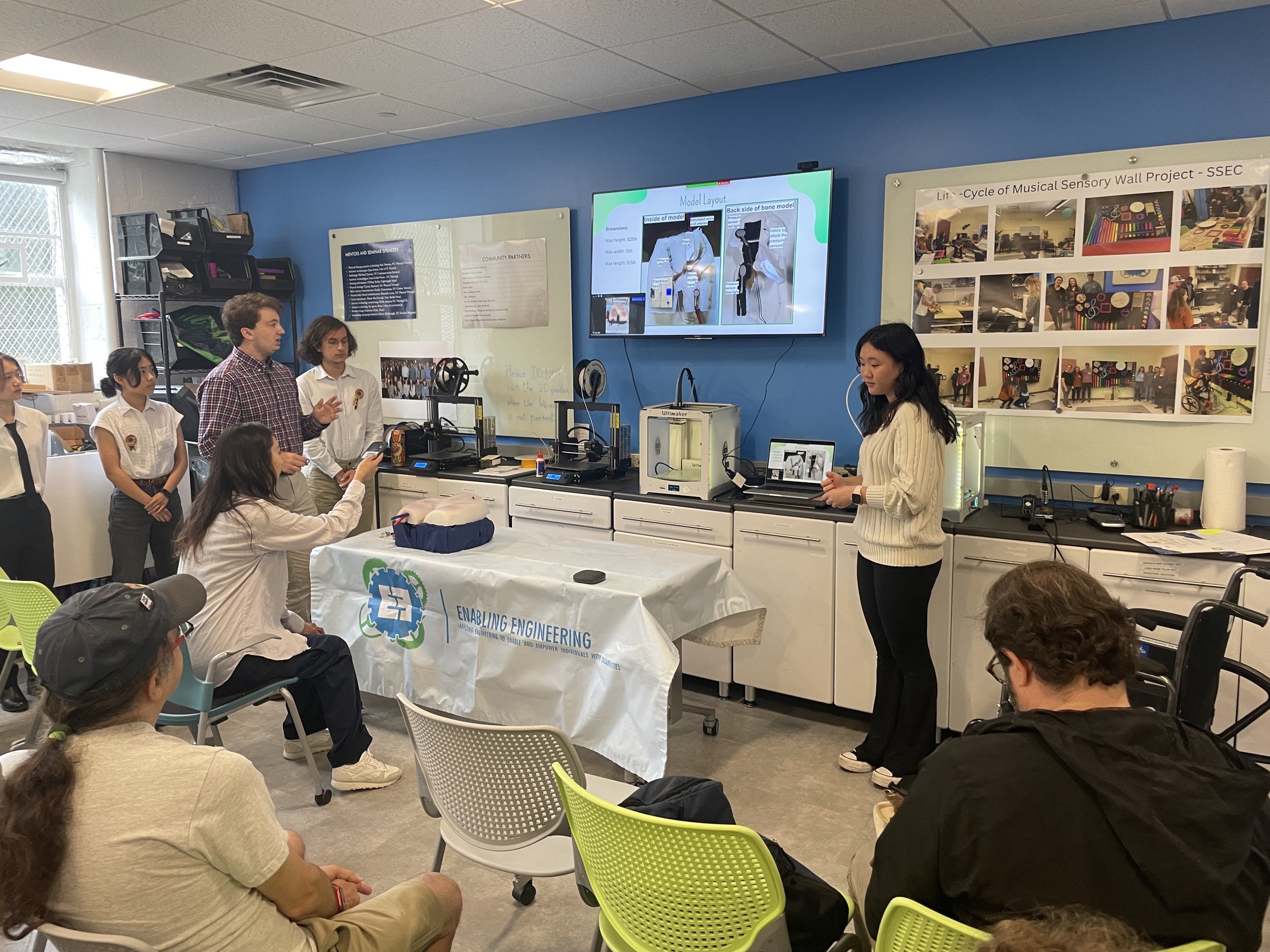
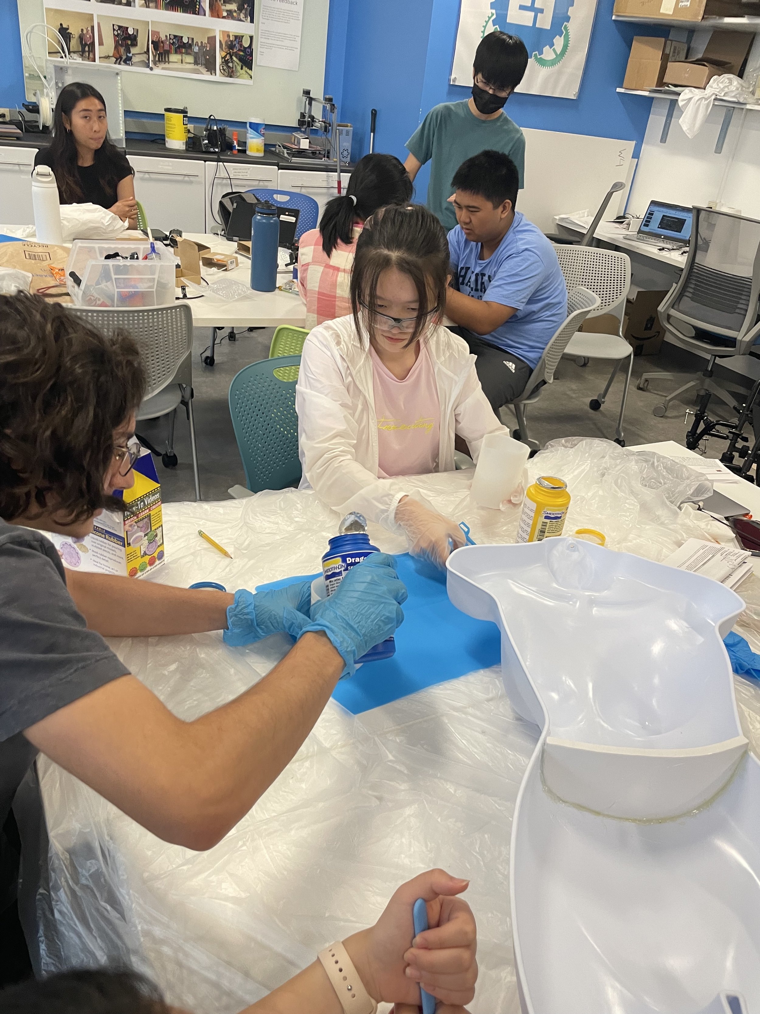
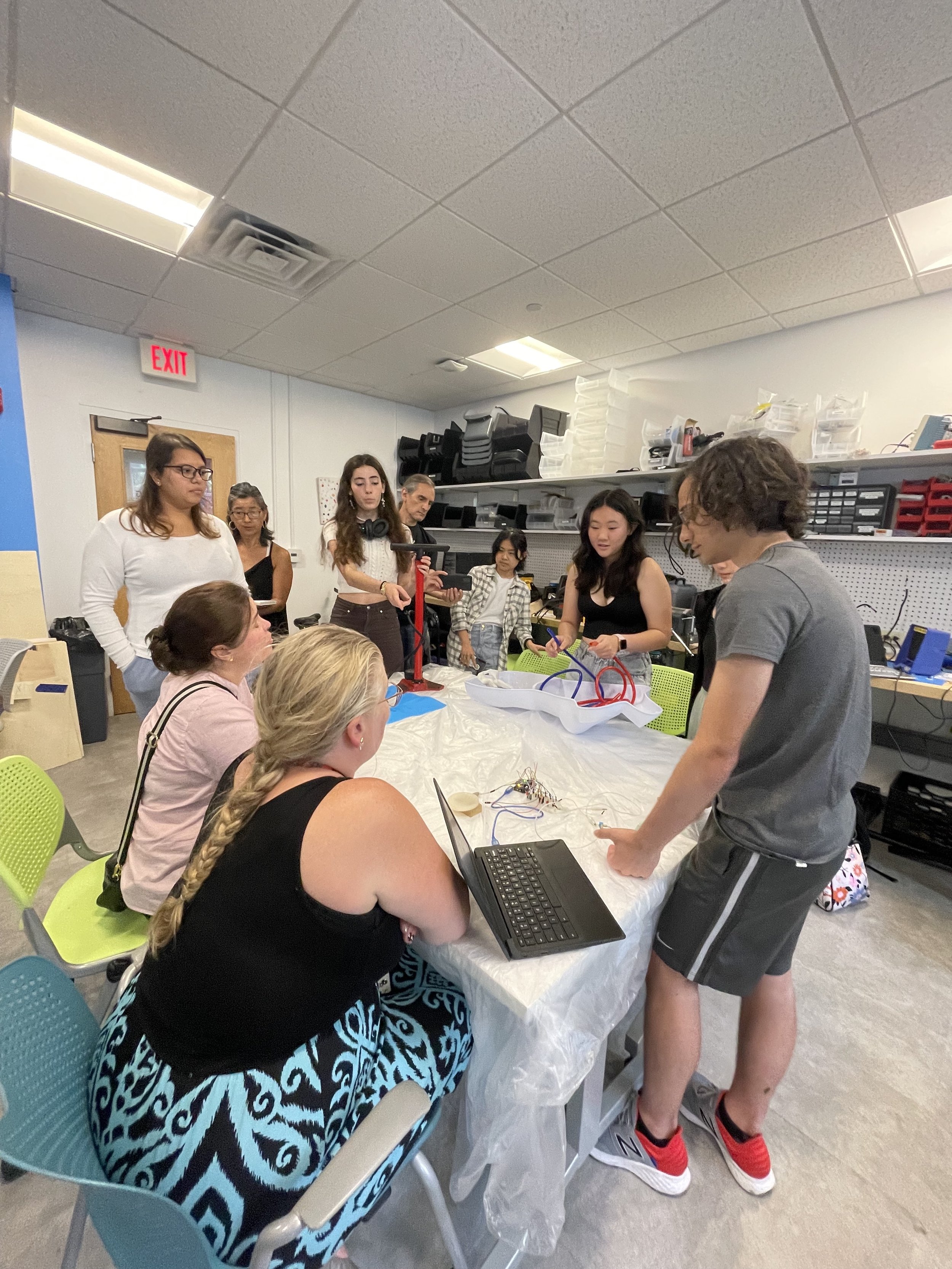
Currently, there are not enough nurses trained to deal with femoral hematomas, and the current training model is neither detailed nor adjustable. We are working on making a model to mimic a femoral hematoma accurately using an inflatable bladder. We will have pressure sensors above and below the simulated hematomas to detect whether the trainee is applying pressure correctly to stop the blood flow. There will also be a pressure sensor below the hematoma to detect if the trainee is using the correct pressure for diffusion. A green and red light will then be lit based on the sensor’s output, green being the correct pressure applied and red for incorrect pressure.
The Team
Isabella Tang
Jacob Freed
Theresa Franklin
Aaron Paker
Dennis Liu
Franklin Ji
Yutong Yan
Lulu Jin